ANATOMY
The Landmarks of the Body
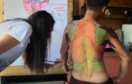
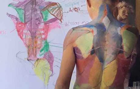
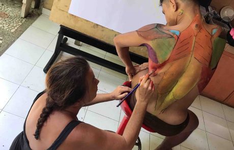
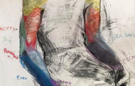
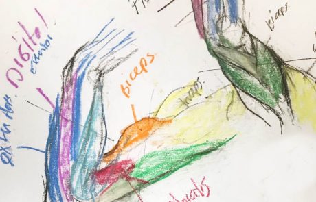
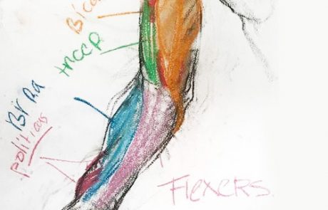
Working with live models gives us the opportunity to carefully study human anatomy. However, I also teach “living anatomy”. This means we focus not so much on the names of the muscles or bones (which are also important for an artist) but also how these change during movement.
When looking at anatomy we have to start with the bones. We have 206 to 213 bones in the human body. The points where bones touch the skin, we call landmarks. These are bony areas that show at the surface. In my drawings, I make these areas darker or sharper, to differentiate shade from muscles. The bone structure of humans is quite similar but the muscles are always different. We need to know the bones because most muscles are attached to the bones.
We work with real-life models because it’s not as easy to locate these landmarks. In anatomy books, they use easier, simple drawings. Some muscles cover up the landmarks, so working with a real-life model means you can search yourself which makes learning this way more interesting. Humans are all different, learning and seeing the difference is learning anatomy. If you’d like to learn drawing anatomy make sure you buy a book that has photos.

This is my Bible, because it also have photos, drawings, starting points of the muscle. You need photos because you need to found it back in the model.
We draw the bones on the model. Most will stay on his place only the shoulder blades will move a lot.
Start to draw a skeleton and find the landmarks on yourself.

Head
- Frontal bone
- Temporal line
- Nasal
- Zygomatic
- Mandible. More corned by male
- Clavicle
Font Body
- Acromion process
- Sternum
- Ribs, Costal Cartilage
- Iliac crest
- ASIS
- Pubic bone
Backside body
- 7Th Cervical Vertebra
- Medial Border of Scapula
- Spine of Scapula
- PSIS
- Sacrum
- Tailbone
Arms and hands
- Olecranon
- Medial Epicondyle of the Humerus
- Ulna Furrow
- Styloid Process of Ulna
- Styloid of process Radius
- Phalanges
Legs
- Greater Trochanter
- Medial Epicondyle of Femur
- Lateral Epicondyle
- Medial condyle of Tibia
- Medial Malleolus
- Lateral Malleolus
- Head of Fibula
Foot
- Phalanges
- Calcaneus
- 5th Metatarsal
Everything where I made red is a landmark= bone close to the skin.


Before we move on to applying structure and proportion to the figure drawings we need to learn the landmarks of the body.
Photos from the classes in December 2018
